
Visit of the Federal Research Minister Prof. Johanna Wanka
 |
| © Uniklinik RWTH Aachen |
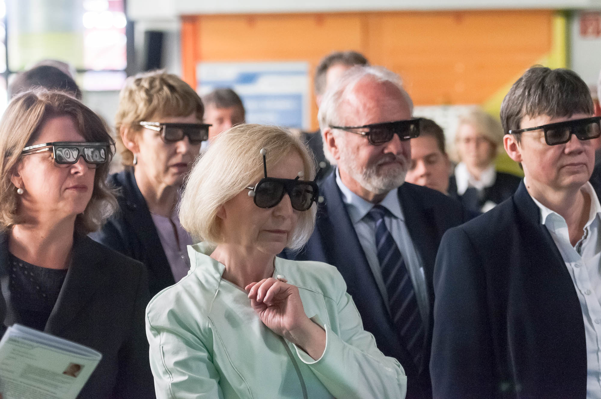
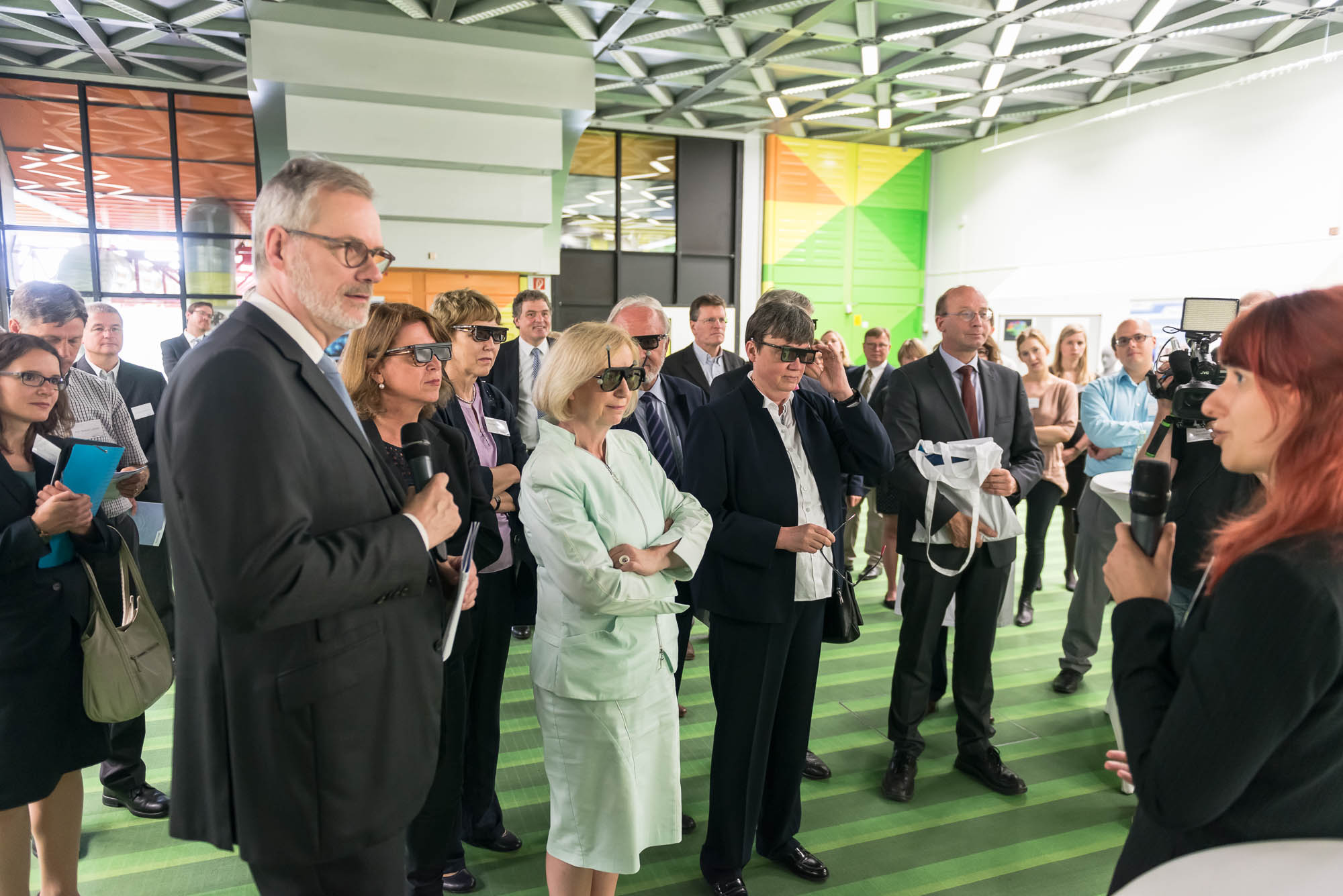
During her summer tour under the topic “The Future of Life in Old Age” Prof. Johanna Wanka visited within the scope of JARA-BRAIN the RWTH Aachen University Hospital on July 28th. Young scientists had the chance to present their research in short talks and demonstrations. For the Virtual Reality Group, Claudia Hänel presented her work on an interactive application to visualize the degenerative progression of a patient’s brain who suffers from corticobasal syndrome. This work was undertaken in a close cooperation with the group of Prof. Katrin Amunts (Institut für Neurowissenschaften und Medizin, Forschungszentrum Jülich) and is currently continued with the scope on the visualization of large data gathered from polarized light imaging. Within her short demonstration on a semi-immersive display system, Claudia Hänel was able to highlight the potential benefits neuroscientists can gain from novel interactive and immersive visualizations of their data.
 |
| © Uniklinik RWTH Aachen |
The visualization of the progression of brain tissue loss in neurodegenerative diseases like corticobasal syndrome can provide not only information about the localization and distribution of the volume loss, but also helps to understand the course and the causes of this neurodegenerative disorder. The visualization of such medical imaging data is often based on 2D sections, because they show both internal and external structures in one image. Spatial information, however, is lost. 3D visualization of imaging data is capable to solve this problem, but it faces the difficulty that more internally located structures may be occluded by structures near the surface. Here, we present an application with two designs for the 3D visualization of the human brain to address these challenges. In the first design, brain anatomy is displayed semi-transparently; it is supplemented by an anatomical section and cortical areas for spatial orientation, and the volumetric data of volume loss. The second design is guided by the principle of importance-driven volume rendering: A direct line-of-sight to the relevant structures in the deeper parts of the brain is provided by cutting out a frustum-like piece of brain tissue. The application was developed to run in both, standard desktop environments and in immersive virtual reality environments with stereoscopic viewing for improving the depth perception. We conclude, that the presented application facilitates the perception of the extent of brain degeneration with respect to its localization and affected regions.
